Double Trouble!!
This 60yo lady had sequential paracentral and central scotomas following trivial trauma and valsalva events.
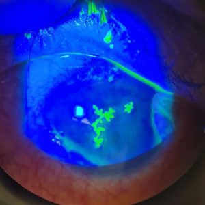
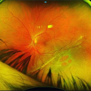
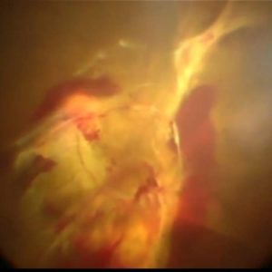
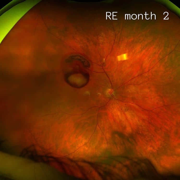
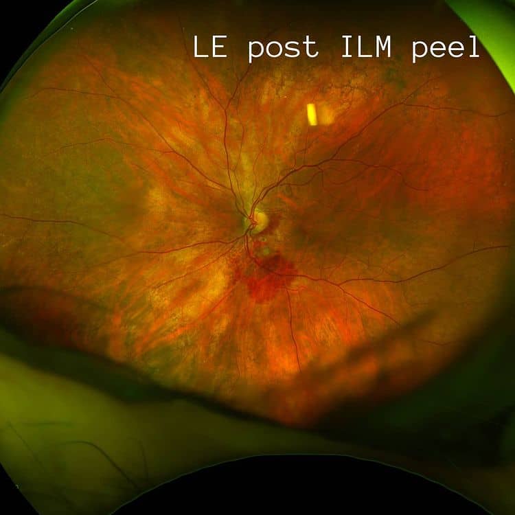
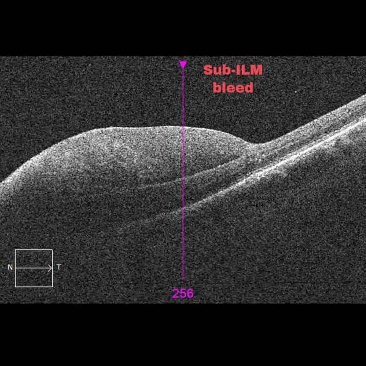
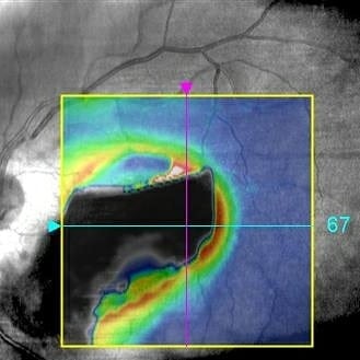
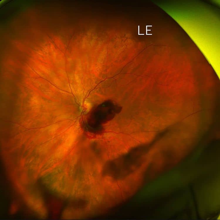
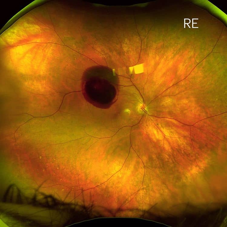
She initially had large multilayered bleeds in both eyes (images 1 &2) and underwent vitrectomy in the left eye for central sub-ILM bleed overlying her macula seen on OCT imaging (images 3&4), fortunately there was no subfoveal blood in either eye.
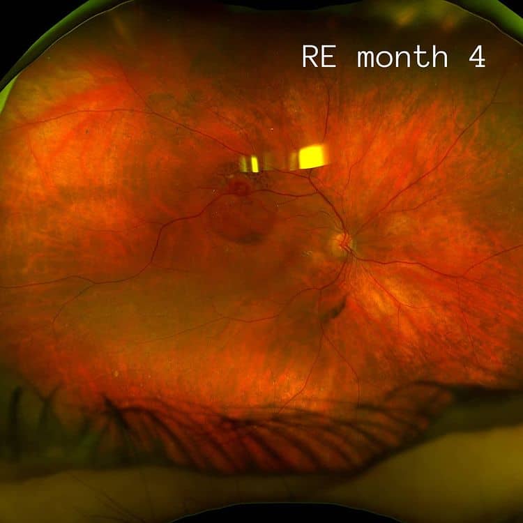
Images 6 & 7 show resolution of the subILM bleed in the RE with observation over 4 months.
She has managed to retain 6/6 vision in both eyes.
These pseudocolour Optos photos show bilateral ruptured Retinal Artery Macroaneurysms (RAMA), most often associated with longstanding systemic hypertension in older females. They are described as a localised dilatation of retinal arteriole within 3 orders of arterial tree at birfurcations, with 90% being unilateral.
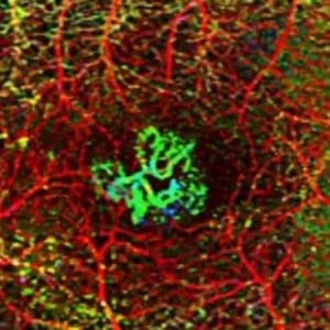
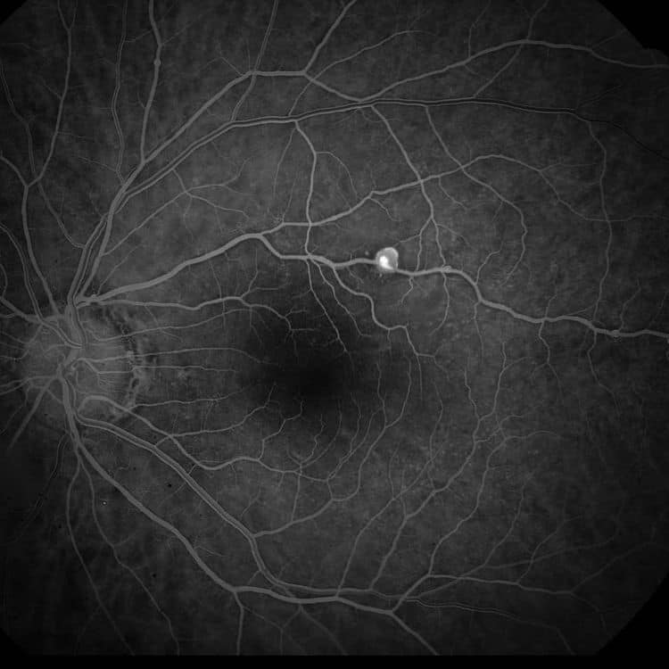
Image 8 is from another patient and demonstrates the typical appearance of a RAMA on angiography.
Treatments usually include observation or thermal laser if there is sight threatening exudation. Surgery is only usually required for ruptured RAMAs that obscure vision through vitreous haemorrhage or central involvement.
It’s important to liaise with the patient’s Primary Care Physician to ensure that cardiovascular risk factors are well controlled.
#retina #ophthalmology #vitreoretinalsurgery #eyedisease #retinasurgery #eyesurgery #ophthalmologist #eyedoctor #optometry #optometrist #optometrystudent #vitrectomy #macula #laser #medicalretina #medret #angiography #optomap #retinopathy #maculopathy #cmo #cme #maculardegeneration #optomap #goldcoast #goldcoasteyes #outlookeye #goldcoasteyesurgeon #goldcoastoptomp


